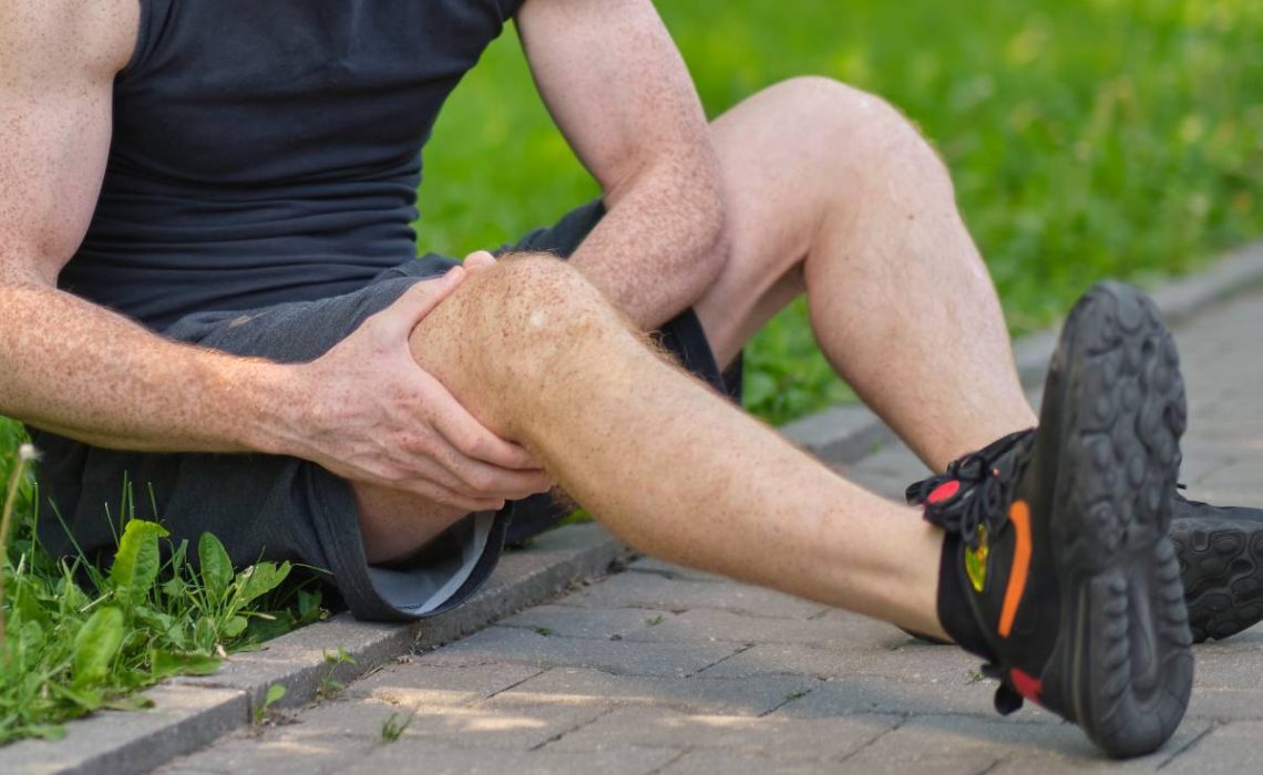
Experiencing mild knee discomfort following a workout is a frequent occurrence, but if it persists, it might indicate a more serious issue.
The knee is a complex joint comprising bones, ligaments, menisci, muscles, and tendons, all of which work together to support movement. If any of these components are damaged or stressed, it can result in achy knees. Various physical activities like bending, jumping, running, and stretching can strain or impact the knees, leading to workout-related pain.
Knee pain is a prevalent concern during exercise, affecting approximately 25% of adults. Keep reading to discover the common reasons behind knee pain and how to effectively address it.
Overuse or strain injuries occur when repetitive stress is placed on a specific body part without sufficient time for recovery. This can result in inflammation, microtears in the tissues, and pain. Here’s an in-depth look at overuse or strain injuries in the context of knee pain after working out:
Mechanisms of Overuse or Strain Injuries
Repetitive Microtrauma: Overuse injuries typically result from repetitive microtrauma to the tissues around the knee. This can include muscles, tendons, ligaments, and bones. Unlike acute injuries caused by a single traumatic event, overuse injuries develop gradually over time due to repeated stress.
Inflammation: Persistent strain can cause inflammation of various structures around the knee:
Tendons (Tendinitis): Tendons are the fibrous tissues that connect muscles to bones. Tendinitis occurs when these tendons become inflamed due to overuse. Common types include patellar tendinitis (jumper’s knee) and quadriceps tendinitis.
Bursae (Bursitis): Bursae are small fluid-filled sacs that reduce friction between tissues. Overuse can cause these sacs to become inflamed, leading to bursitis. Prepatellar bursitis (housemaid’s knee) is a common example.
Tissue Breakdown: Continuous overuse without adequate rest can lead to the breakdown of tissues faster than the body can repair them. This can result in microtears in the tendons and muscles, leading to pain and dysfunction.
Contributing Factors
High-Impact Activities: Activities that involve repetitive high-impact movements, such as running, jumping, and certain sports, can increase the risk of overuse injuries.
Training Errors: Rapid increases in the intensity, duration, or frequency of workouts can overload the knee joint and surrounding tissues.
Poor Biomechanics: Abnormal movement patterns, improper alignment, or imbalances in muscle strength and flexibility can contribute to increased strain on the knee.
Inadequate Recovery: Not allowing enough time for rest and recovery between workouts can prevent tissues from healing
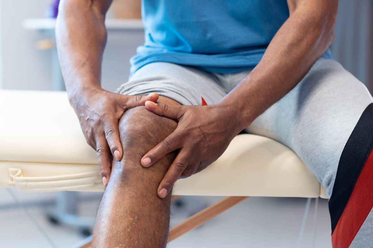
Possible cause no.2: Arthritis
Arthritis is a common cause of knee pain, especially after physical activity. It refers to inflammation of one or more joints, which can lead to pain, swelling, stiffness, and decreased range of motion. There are several types of arthritis that can affect the knee, with osteoarthritis being the most prevalent. Here’s a detailed look at arthritis in the context of knee pain:
Types of Arthritis Affecting the Knee
Osteoarthritis (OA):
Description: Osteoarthritis is a degenerative joint disease characterized by the breakdown of cartilage, the smooth tissue that cushions the ends of bones in the joint. As the cartilage wears away, bones can rub against each other, causing pain, swelling, and stiffness.
Causes: It can be caused by ageing, joint injury, repetitive stress on the joint, and genetic factors. Overweight and obesity also increase the risk as they put additional stress on the knee joints.
Symptoms: Pain that worsens with activity and improves with rest, stiffness (especially after periods of inactivity), swelling, and a reduced range of motion. Patients may also experience a grinding sensation (crepitus) during joint movement.
Rheumatoid Arthritis (RA):
Description: Rheumatoid arthritis is an autoimmune disease in which the immune system mistakenly attacks the synovium (the lining of the membranes that surround the joints), leading to inflammation and joint damage.
Causes: The exact cause is unknown, but it involves genetic, environmental, and hormonal factors. Smoking and certain infections have been linked to the onset of RA.
Symptoms: Joint pain, swelling, and stiffness, often affecting both knees symmetrically. Systemic symptoms such as fatigue, fever, and loss of appetite may also be present. Morning stiffness lasting more than 30 minutes is common.
Post-Traumatic Arthritis:
Description: This type of arthritis develops after an injury to the knee, such as a fracture, ligament injury, or meniscus tear. The injury can lead to joint instability, altered joint mechanics, and eventual cartilage breakdown.
Causes: Knee injuries that were not adequately treated or healed improperly can lead to post-traumatic arthritis.
Symptoms: Pain, swelling, and stiffness, which may develop months or years after the initial injury.
Gout and Pseudogout:
Gout: Caused by the accumulation of uric acid crystals in the joint, leading to sudden and severe pain, redness, and swelling.
Pseudogout: Caused by calcium pyrophosphate crystal deposition in the joint. Symptoms are similar to gout but usually less severe.
Pathophysiology
Inflammation: In arthritis, the affected joint becomes inflamed. In OA, this is due to mechanical wear and tear, while in RA, it’s due to immune system activity.
Cartilage Degradation: The cartilage in the knee joint breaks down, leading to bone-on-bone contact. This process is gradual in OA and can be accelerated in post-traumatic arthritis.
Bone Changes: As the cartilage deteriorates, bones may develop growths called osteophytes (bone spurs), which can cause additional pain and restrict joint movement.
Synovial Changes: In RA, the synovial membrane becomes thickened and produces excess synovial fluid, causing swelling and pain.
Diagnosis
Clinical Examination: Assessment of symptoms, joint examination for swelling, tenderness, and range of motion.
Imaging: X-rays to detect joint space narrowing, osteophytes, and bone changes. MRI and ultrasound can provide detailed images of soft tissues and detect early changes in RA.
Laboratory Tests: Blood tests for inflammatory markers (CRP, ESR), rheumatoid factor (RF), anti-CCP antibodies (specific for RA), and uric acid levels (for gout).
Management and Treatment
Medications:
Analgesics: Pain relievers like acetaminophen.
NSAIDs: Nonsteroidal anti-inflammatory drugs (ibuprofen, naproxen) reduce pain and inflammation.
Corticosteroids: Oral or injectable steroids to reduce inflammation.
DMARDs: Disease-modifying antirheumatic drugs (methotrexate, sulfasalazine) for RA.
Biologics: Targeted therapy for RA (TNF inhibitors, IL-6 inhibitors).
Physical Therapy:
Exercises to strengthen muscles around the knee and improve flexibility.
Techniques to improve joint function and reduce pain.
Lifestyle Modifications:
Weight management to reduce stress on the knee joint.
Low-impact exercises such as swimming or cycling.
Assistive Devices:
Braces or orthotics to support the knee.
Use of canes or walkers if necessary.
Surgical Options:
Arthroscopy: Minimally invasive surgery to remove loose cartilage or repair damaged tissues.
Osteotomy: Bone cutting procedure to realign the knee joint.
Joint Replacement: Partial or total knee replacement for severe arthritis.
Preventive Measures
Regular Exercise: Strengthening and flexibility exercises to support joint health.
Proper Technique: Using correct form during activities to avoid undue stress on the knee.
Injury Prevention: Protective gear and safe practices to prevent knee injuries.
Healthy Diet: Nutrient-rich diet to maintain joint and overall health.
Understanding arthritis in-depth helps in recognizing the symptoms early and adopting appropriate measures to manage the condition effectively and improve the quality of life.
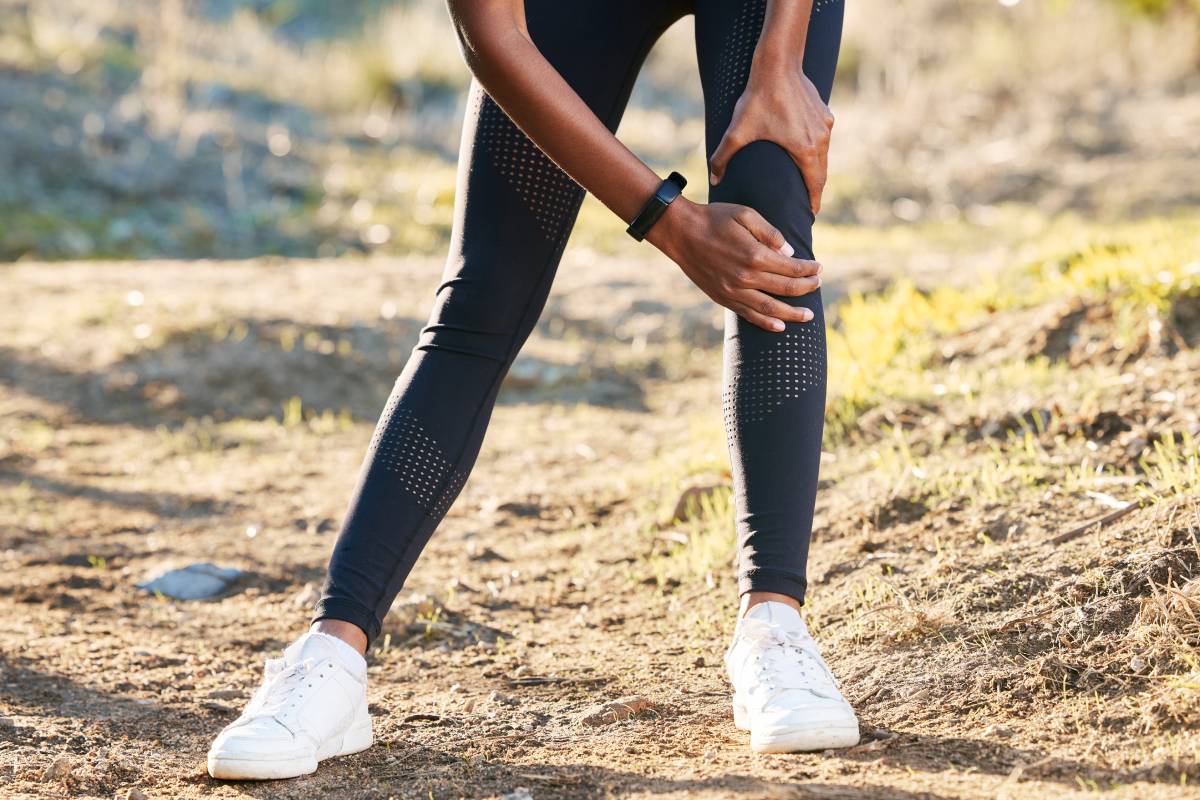
Possible cause no.3: Patellofemoral Pain Syndrome
Patellofemoral Pain Syndrome (PFPS), also known as “runner’s knee,” is a common condition characterized by pain around or behind the patella (kneecap). It is particularly prevalent among athletes, adolescents, and those engaged in repetitive knee-stressing activities. Here’s a detailed look at PFPS:
Causes and Risk Factors
Overuse: Repetitive activities that put stress on the knee, such as running, jumping, and squatting, can lead to PFPS. The repetitive motion can irritate the patellofemoral joint.
Biomechanical Issues: Abnormal movement patterns can increase stress on the knee:
Malalignment: If the patella does not track properly in the femoral groove, it can cause pain. Factors contributing to malalignment include a high-riding patella (patella alta), flat feet (pes planus), or excessive inward rotation of the femur (femoral anteversion).
Muscle Imbalance: Weakness or imbalance in the quadriceps, particularly the vastus medialis obliquus (VMO), can lead to improper patellar tracking.
Tightness: Tightness in the iliotibial band (IT band), hamstrings, or calf muscles can alter patellar tracking.
Trauma: A direct blow to the kneecap or a sudden twist can precipitate PFPS.
Inadequate Footwear: Worn-out or inappropriate shoes that do not provide proper support can exacerbate knee pain.
Symptoms
Anterior Knee Pain: Pain is usually located around or behind the kneecap, often described as a dull, aching pain.
Pain During Activities: Activities such as running, climbing stairs, squatting, or sitting with bent knees for long periods (theatre sign) can exacerbate the pain.
Crepitus: A grinding or cracking sensation in the knee during movement.
Swelling: Mild swelling may occur in the knee joint.
Giving Way: Occasionally, the knee may feel like it’s giving out, due to pain or muscle inhibition.
Diagnosis
Clinical Examination: A thorough history and physical examination to assess pain location, knee alignment, muscle strength, and flexibility. Specific tests may include:
Patellar Apprehension Test: To check for patellar instability.
Clark’s Sign (Patellar Grind Test): Applying pressure to the patella while the patient contracts the quadriceps.
Imaging: Usually not necessary for diagnosis but may be used to rule out other conditions.
X-rays: To check for patellar alignment and rule out bone abnormalities.
MRI: In cases where soft tissue or cartilage damage is suspected.
Treatment
Rest and Activity Modification: Reducing or avoiding activities that exacerbate the pain. Incorporating low-impact exercises such as swimming or cycling.
Physical Therapy: Focuses on:
Strengthening Exercises: To improve the strength of the quadriceps, especially the VMO, and other supporting muscles (hip abductors and external rotators).
Stretching: For the IT band, hamstrings, and calf muscles to improve flexibility.
Patellar Taping or Bracing: To improve patellar tracking and reduce pain.
Footwear: Wearing proper shoes with good arch support. Orthotic inserts may be recommended for those with flat feet.
Pain Management:
NSAIDs: Nonsteroidal anti-inflammatory drugs like ibuprofen to reduce pain and inflammation.
Ice Therapy: Applying ice packs to the knee to manage pain and swelling.
Activity Modification and Gradual Return to Activity: Modifying activities to avoid pain and gradually increasing intensity as symptoms improve.
Preventive Measures
Strength Training: Regularly performing exercises to strengthen the quadriceps, hamstrings, glutes, and hip muscles to maintain balanced muscle strength around the knee.
Flexibility Exercises: Stretching the lower extremity muscles, including the quadriceps, hamstrings, and IT band, to maintain flexibility.
Proper Footwear: Wear appropriate shoes for specific activities and replace them regularly to ensure they provide adequate support.
Technique Optimization: Ensuring proper form and technique during physical activities to minimize stress on the knee.
Gradual Progression: Slowly increasing the intensity and duration of physical activities to allow the body to adapt and prevent overuse injuries.
Long-Term Management
Education: Understanding the condition and learning strategies to manage symptoms can help prevent recurrence.
Consistency with Exercise: Regular adherence to strengthening and stretching routines is essential for long-term knee health.
Monitoring Symptoms: Keeping track of symptoms and activity levels to identify patterns and make adjustments as needed.
Patellofemoral Pain Syndrome is a manageable condition with appropriate interventions focusing on reducing pain, improving knee function, and preventing future episodes.
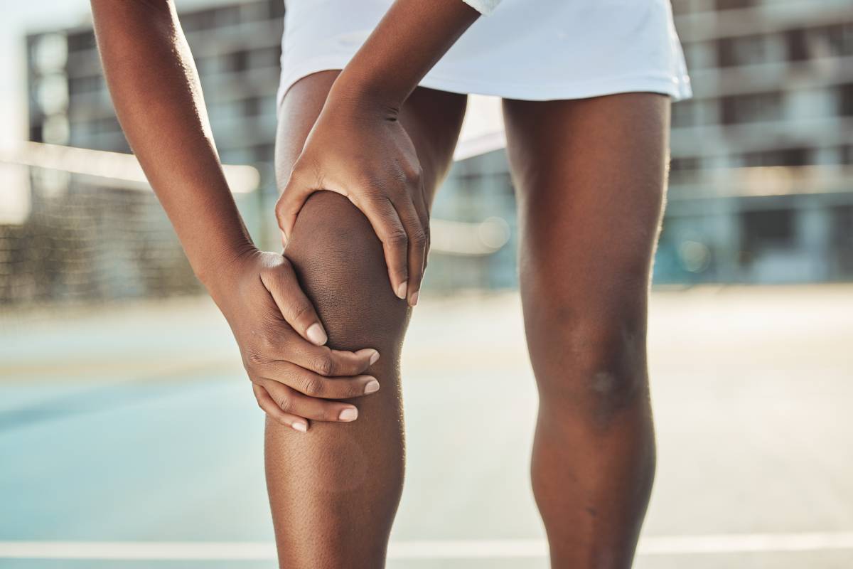
Possible cause no.4: IT Band Syndrome
Iliotibial Band Syndrome (ITBS), also known as IT Band Syndrome, is a common overuse injury affecting the outer part of the knee, often seen in runners, cyclists, and athletes who engage in repetitive knee-bending activities. Here’s an in-depth look at ITBS:
Anatomy and Function of the IT Band
The iliotibial (IT) band is a thick band of fibrous tissue that runs along the outside of the thigh, from the hip (ilium) to just below the knee (tibia). It functions to:
Stabilize the Knee: Provides lateral stability to the knee during movement.
Assist in Movement: Works with the thigh muscles to extend, abduct, and rotate the hip, and to stabilize the knee during running or walking.
Causes and Risk Factors
Overuse and Repetitive Activity: Engaging in activities that involve repetitive knee flexion and extension, such as running, cycling, and hiking, can lead to overuse of the IT band.
Biomechanical Factors:
Leg Length Discrepancy: One leg being longer than the other can cause an imbalance, increasing stress on the IT band.
Foot Mechanics: Overpronation (excessive inward rolling of the foot) or supination (outward rolling) can affect knee alignment and increase IT band strain.
Hip Muscle Weakness: Weak hip abductors (gluteus medius) can cause the pelvis to drop on the opposite side during walking or running, increasing tension on the IT band.
Training Errors: Sudden increases in training intensity, duration, or frequency can overload the IT band. Running on sloped or uneven surfaces can also contribute.
Improper Footwear: Worn-out or inappropriate shoes can lead to poor foot mechanics and increase stress on the IT band.
Poor Running Technique: Running with improper form, such as excessive stride length or running on one side of a cambered road, can exacerbate IT band issues.
Symptoms
Lateral Knee Pain: The primary symptom is pain on the outside of the knee, which may start as an ache and progress to a sharp or burning sensation.
Pain During Activity: Pain typically worsens with activities involving knee flexion and extension, such as running (especially downhill), cycling, or climbing stairs.
Tenderness: Tenderness to touch over the lateral femoral epicondyle (the bony prominence on the outer knee).
Snapping Sensation: Some individuals may experience a snapping or popping sensation on the outside of the knee.
Diagnosis
Clinical Examination: A healthcare provider will assess the patient’s medical history, symptoms, and conduct a physical examination focusing on the knee and hip alignment, muscle strength, and flexibility. Key tests include:
Noble’s Compression Test: Applying pressure to the IT band just above the knee while the patient flexes and extends the knee. Pain at about 30 degrees of knee flexion is indicative of ITBS.
Ober’s Test: Assessing IT band tightness by having the patient lie on their side, extending the hip and allowing the leg to drop towards the table. Inability to lower the leg smoothly suggests IT band tightness.
Imaging: Generally not required for diagnosis but may be used to rule out other conditions. MRI can show inflammation or thickening of the IT band.
Treatment
Rest and Activity Modification: Reducing or avoiding activities that aggravate the pain to allow the IT band to heal. Cross-training with low-impact activities like swimming can be beneficial.
Physical Therapy:
Stretching: Regular stretching exercises for the IT band, hamstrings, quadriceps, and hip muscles.
Strengthening: Strengthening exercises for the hip abductors, gluteal muscles, and core to improve stability and reduce strain on the IT band.
Foam Rolling: Using a foam roller to massage and release tension in the IT band and surrounding muscles.
Pain Management:
NSAIDs: Nonsteroidal anti-inflammatory drugs like ibuprofen or naproxen to reduce pain and inflammation.
Ice Therapy: Applying ice to the affected area to reduce pain and swelling.
Proper Footwear: Ensuring shoes provide adequate support and cushioning. Orthotic inserts may be recommended for those with foot alignment issues.
Running Form: Addressing running mechanics and technique with a coach or physical therapist to avoid exacerbating the condition.
Prevention
Gradual Progression: Gradually increasing the intensity and duration of activities to allow the body to adapt.
Strength Training: Regularly performing exercises to strengthen the hip, gluteal, and core muscles to maintain stability and support.
Flexibility Exercises: Incorporating stretches for the IT band, quadriceps, hamstrings, and calves to maintain flexibility.
Proper Equipment: Using appropriate footwear for specific activities and replacing shoes regularly to ensure they provide proper support.
Surface Variation: Avoiding repetitive activities on sloped or uneven surfaces, and varying running routes to prevent overloading one side of the body.
Long-Term Management
Consistency with Exercises: Regularly performing strength and flexibility exercises to prevent recurrence.
Monitoring Symptoms: Keeping track of symptoms and activity levels to identify triggers and make necessary adjustments.
Professional Guidance: Working with healthcare professionals, such as physical therapists, to develop and maintain an effective management plan.
Understanding IT Band Syndrome and adopting appropriate measures can help manage symptoms, prevent recurrence, and ensure a quicker return to pain-free activity.
Possible cause no.5: Improper Footwear
Improper footwear can contribute significantly to various lower limb injuries and conditions, including knee pain. Here’s a detailed look at how inappropriate footwear can affect knee health:
Impact on Knee Health
Lack of Support: Shoes with inadequate support fail to stabilize the foot and ankle properly, leading to poor biomechanics. This can result in altered gait patterns, placing excessive stress on the knee joint during weight-bearing activities.
Poor Cushioning: Shoes with insufficient cushioning offer little shock absorption, leading to increased impact forces transmitted to the knee joint. Over time, this can contribute to joint pain and discomfort, especially during high-impact activities like running or jumping.
Improper Alignment: Footwear that does not provide proper alignment for the foot and ankle can affect lower limb alignment, leading to overpronation (excessive inward rolling of the foot) or supination (outward rolling). These abnormal movement patterns can alter knee biomechanics and increase the risk of injuries such as IT band syndrome, patellofemoral pain syndrome, and runner’s knee.
Characteristics of Proper Footwear
Arch Support: Shoes with adequate arch support help maintain the natural arch of the foot, providing stability and reducing the risk of overpronation.
Cushioning: Good cushioning in the midsole and heel absorbs shock during impact, reducing stress on the knee joint and surrounding structures.
Stability Features: Shoes with features like a firm heel counter and supportive upper help stabilize the foot and ankle, promoting proper alignment and reducing the risk of injury.
Proper Fit: Shoes should fit comfortably with enough room for the toes to wiggle but without excessive movement within the shoe. Poorly fitting shoes can lead to blisters, calluses, and foot deformities that can indirectly affect knee health.
Activity-Specific Design: Different activities require specific footwear designs to accommodate the unique demands of the sport or activity. For example, running shoes typically have more cushioning and flexibility, while hiking boots offer greater ankle support and traction.
Common Types of Improper Footwear
Worn-Out Shoes: Shoes that are past their prime lose their supportive and cushioning properties, increasing the risk of knee pain and injury.
Fashion Footwear: Fashionable shoes often prioritize style over function, lacking proper support and cushioning. Prolonged wear of such shoes can contribute to foot and knee problems.
Inappropriate Shoes for Activity: Using shoes not designed for a specific activity, such as wearing running shoes for hiking or basketball shoes for tennis, can lead to inadequate support and increased injury risk.
High Heels: High-heeled shoes alter lower limb biomechanics, shifting the body’s weight forward and increasing stress on the knees. Prolonged wear of high heels can contribute to knee pain and musculoskeletal imbalances.
Flat Shoes: Shoes with insufficient arch support and cushioning, such as flip-flops or certain types of sandals, provide little to no shock absorption and can exacerbate knee pain, especially during prolonged walking or standing.
Tips for Choosing Proper Footwear
Get Properly Fitted: Visit a reputable shoe store where knowledgeable staff can measure your feet and recommend suitable footwear based on your foot shape and activity level.
Consider Activity-Specific Shoes: Invest in shoes designed for the activities you participate in regularly, whether it’s running, walking, hiking, or playing sports.
Replace Worn-Out Shoes: Monitor the condition of your shoes and replace them when signs of wear and tear, such as worn-out treads or flattened cushioning, become apparent.
Prioritize Comfort and Function: Choose shoes that feel comfortable and supportive from the moment you try them on. Avoid sacrificing comfort for style.
Orthotic Inserts: If you have specific foot issues or biomechanical imbalances, consider using orthotic inserts or custom orthotics prescribed by a podiatrist to provide additional support and stability.
When to Seek Medical Attention
If you experience severe knee pain, it’s important to consult a healthcare provider promptly. This could indicate a serious knee condition, such as a dislocated or fractured kneecap.
Other signs that warrant a visit to a healthcare provider include:
Buckling, clicking, or locking sensations in the knee
Deformity or noticeable change in the shape of the knee
Presence of fever, redness, or warmth accompanied by swelling in the knee area
Persistence of pain even after three days of self-care measures at home
Difficulty in flexing or straightening the knee
Treatment approaches for severe knee pain can vary. Depending on the underlying condition, a healthcare provider may administer a steroid injection to alleviate pain and swelling. They might also recommend referral to a physical therapist for further management. In cases of severe knee conditions, surgical intervention may be necessary.
Patellofemoral Pain Syndrome (PFPS), also known as “runner’s knee,” is a common condition characterized by pain around or behind the patella (kneecap). It is particularly prevalent among athletes, adolescents, and those engaged in repetitive knee-stressing activities. Here’s a detailed look at PFPS:
Causes and Risk Factors
Overuse: Repetitive activities that put stress on the knee, such as running, jumping, and squatting, can lead to PFPS. The repetitive motion can irritate the patellofemoral joint.
Biomechanical Issues: Abnormal movement patterns can increase stress on the knee:
Malalignment: If the patella does not track properly in the femoral groove, it can cause pain. Factors contributing to malalignment include a high-riding patella (patella alta), flat feet (pes planus), or excessive inward rotation of the femur (femoral anteversion).
Muscle Imbalance: Weakness or imbalance in the quadriceps, particularly the vastus medialis obliquus (VMO), can lead to improper patellar tracking.
Tightness: Tightness in the iliotibial band (IT band), hamstrings, or calf muscles can alter patellar tracking.
Trauma: A direct blow to the kneecap or a sudden twist can precipitate PFPS.
Inadequate Footwear: Worn-out or inappropriate shoes that do not provide proper support can exacerbate knee pain.
Symptoms
Anterior Knee Pain: Pain is usually located around or behind the kneecap, often described as a dull, aching pain.
Pain During Activities: Activities such as running, climbing stairs, squatting, or sitting with bent knees for long periods (theatre sign) can exacerbate the pain.
Crepitus: A grinding or cracking sensation in the knee during movement.
Swelling: Mild swelling may occur in the knee joint.
Giving Way: Occasionally, the knee may feel like it’s giving out, due to pain or muscle inhibition.
Diagnosis
Clinical Examination: A thorough history and physical examination to assess pain location, knee alignment, muscle strength, and flexibility. Specific tests may include:
Patellar Apprehension Test: To check for patellar instability.
Clark’s Sign (Patellar Grind Test): Applying pressure to the patella while the patient contracts the quadriceps.
Imaging: Usually not necessary for diagnosis but may be used to rule out other conditions.
X-rays: To check for patellar alignment and rule out bone abnormalities.
MRI: In cases where soft tissue or cartilage damage is suspected.
Treatment
Rest and Activity Modification: Reducing or avoiding activities that exacerbate the pain. Incorporating low-impact exercises such as swimming or cycling.
Physical Therapy: Focuses on:
Strengthening Exercises: To improve the strength of the quadriceps, especially the VMO, and other supporting muscles (hip abductors and external rotators).
Stretching: For the IT band, hamstrings, and calf muscles to improve flexibility.
Patellar Taping or Bracing: To improve patellar tracking and reduce pain.
Footwear: Wearing proper shoes with good arch support. Orthotic inserts may be recommended for those with flat feet.
Pain Management:
NSAIDs: Nonsteroidal anti-inflammatory drugs like ibuprofen to reduce pain and inflammation.
Ice Therapy: Applying ice packs to the knee to manage pain and swelling.
Activity Modification and Gradual Return to Activity: Modifying activities to avoid pain and gradually increasing intensity as symptoms improve.
Preventive Measures
Strength Training: Regularly performing exercises to strengthen the quadriceps, hamstrings, glutes, and hip muscles to maintain balanced muscle strength around the knee.
Flexibility Exercises: Stretching the lower extremity muscles, including the quadriceps, hamstrings, and IT band, to maintain flexibility.
Proper Footwear: Wearing appropriate shoes for specific activities and replace them regularly to ensure they provide adequate support.
Technique Optimization: Ensuring proper form and technique during physical activities to minimize stress on the knee.
Gradual Progression: Slowly increasing the intensity and duration of physical activities to allow the body to adapt and prevent overuse injuries.
Long-Term Management
Education: Understanding the condition and learning strategies to manage symptoms can help prevent recurrence.
Consistency with Exercise: Regular adherence to strengthening and stretching routines is essential for long-term knee health.
Monitoring Symptoms: Keeping track of symptoms and activity levels to identify patterns and make adjustments as needed.
Patellofemoral Pain Syndrome is a manageable condition with appropriate interventions focusing on reducing pain, improving knee function, and preventing future episodes.
Possible cause no.4: IT Band Syndrome
Iliotibial Band Syndrome (ITBS), also known as IT Band Syndrome, is a common overuse injury affecting the outer part of the knee, often seen in runners, cyclists, and athletes who engage in repetitive knee-bending activities. Here’s an in-depth look at ITBS:
Anatomy and Function of the IT Band
The iliotibial (IT) band is a thick band of fibrous tissue that runs along the outside of the thigh, from the hip (ilium) to just below the knee (tibia). It functions to:
Stabilize the Knee: Provides lateral stability to the knee during movement.
Assist in Movement: Works with the thigh muscles to extend, abduct, and rotate the hip, and to stabilize the knee during running or walking.
Causes and Risk Factors
Overuse and Repetitive Activity: Engaging in activities that involve repetitive knee flexion and extension, such as running, cycling, and hiking, can lead to overuse of the IT band.
Biomechanical Factors:
Leg Length Discrepancy: One leg being longer than the other can cause an imbalance, increasing stress on the IT band.
Foot Mechanics: Overpronation (excessive inward rolling of the foot) or supination (outward rolling) can affect knee alignment and increase IT band strain.
Hip Muscle Weakness: Weak hip abductors (gluteus medius) can cause the pelvis to drop on the opposite side during walking or running, increasing tension on the IT band.
Training Errors: Sudden increases in training intensity, duration, or frequency can overload the IT band. Running on sloped or uneven surfaces can also contribute.
Improper Footwear: Worn-out or inappropriate shoes can lead to poor foot mechanics and increase stress on the IT band.
Poor Running Technique: Running with improper form, such as excessive stride length or running on one side of a cambered road, can exacerbate IT band issues.
Symptoms
Lateral Knee Pain: The primary symptom is pain on the outside of the knee, which may start as an ache and progress to a sharp or burning sensation.
Pain During Activity: Pain typically worsens with activities involving knee flexion and extension, such as running (especially downhill), cycling, or climbing stairs.
Tenderness: Tenderness to touch over the lateral femoral epicondyle (the bony prominence on the outer knee).
Snapping Sensation: Some individuals may experience a snapping or popping sensation on the outside of the knee.
Diagnosis
Clinical Examination: A healthcare provider will assess the patient’s medical history, symptoms, and conduct a physical examination focusing on the knee and hip alignment, muscle strength, and flexibility. Key tests include:
Noble’s Compression Test: Applying pressure to the IT band just above the knee while the patient flexes and extends the knee. Pain at about 30 degrees of knee flexion is indicative of ITBS.
Ober’s Test: Assessing IT band tightness by having the patient lie on their side, extending the hip and allowing the leg to drop towards the table. Inability to lower the leg smoothly suggests IT band tightness.
Imaging: Generally not required for diagnosis but may be used to rule out other conditions. MRI can show inflammation or thickening of the IT band.
Treatment
Rest and Activity Modification: Reducing or avoiding activities that aggravate the pain to allow the IT band to heal. Cross-training with low-impact activities like swimming can be beneficial.
Physical Therapy:
Stretching: Regular stretching exercises for the IT band, hamstrings, quadriceps, and hip muscles.
Strengthening: Strengthening exercises for the hip abductors, gluteal muscles, and core to improve stability and reduce strain on the IT band.
Foam Rolling: Using a foam roller to massage and release tension in the IT band and surrounding muscles.
Pain Management:
NSAIDs: Nonsteroidal anti-inflammatory drugs like ibuprofen or naproxen to reduce pain and inflammation.
Ice Therapy: Applying ice to the affected area to reduce pain and swelling.
Proper Footwear: Ensuring shoes provide adequate support and cushioning. Orthotic inserts may be recommended for those with foot alignment issues.
Running Form: Addressing running mechanics and technique with a coach or physical therapist to avoid exacerbating the condition.
Prevention
Gradual Progression: Gradually increasing the intensity and duration of activities to allow the body to adapt.
Strength Training: Regularly performing exercises to strengthen the hip, gluteal, and core muscles to maintain stability and support.
Flexibility Exercises: Incorporating stretches for the IT band, quadriceps, hamstrings, and calves to maintain flexibility.
Proper Equipment: Using appropriate footwear for specific activities and replacing shoes regularly to ensure they provide proper support.
Surface Variation: Avoiding repetitive activities on sloped or uneven surfaces, and varying running routes to prevent overloading one side of the body.
Long-Term Management
Consistency with Exercises: Regularly performing strength and flexibility exercises to prevent recurrence.
Monitoring Symptoms: Keeping track of symptoms and activity levels to identify triggers and make necessary adjustments.
Professional Guidance: Working with healthcare professionals, such as physical therapists, to develop and maintain an effective management plan.
Understanding IT Band Syndrome and adopting appropriate measures can help manage symptoms, prevent recurrence, and ensure a quicker return to pain-free activity.
Possible cause no.5: Improper Footwear
Improper footwear can contribute significantly to various lower limb injuries and conditions, including knee pain. Here’s a detailed look at how inappropriate footwear can affect knee health:
Impact on Knee Health
Lack of Support: Shoes with inadequate support fail to stabilize the foot and ankle properly, leading to poor biomechanics. This can result in altered gait patterns, placing excessive stress on the knee joint during weight-bearing activities.
Poor Cushioning: Shoes with insufficient cushioning offer little shock absorption, leading to increased impact forces transmitted to the knee joint. Over time, this can contribute to joint pain and discomfort, especially during high-impact activities like running or jumping.
Improper Alignment: Footwear that does not provide proper alignment for the foot and ankle can affect lower limb alignment, leading to overpronation (excessive inward rolling of the foot) or supination (outward rolling). These abnormal movement patterns can alter knee biomechanics and increase the risk of injuries such as IT band syndrome, patellofemoral pain syndrome, and runner’s knee.
Characteristics of Proper Footwear
Arch Support: Shoes with adequate arch support help maintain the natural arch of the foot, providing stability and reducing the risk of overpronation.
Cushioning: Good cushioning in the midsole and heel absorbs shock during impact, reducing stress on the knee joint and surrounding structures.
Stability Features: Shoes with features like a firm heel counter and supportive upper help stabilize the foot and ankle, promoting proper alignment and reducing the risk of injury.
Proper Fit: Shoes should fit comfortably with enough room for the toes to wiggle but without excessive movement within the shoe. Poorly fitting shoes can lead to blisters, calluses, and foot deformities that can indirectly affect knee health.
Activity-Specific Design: Different activities require specific footwear designs to accommodate the unique demands of the sport or activity. For example, running shoes typically have more cushioning and flexibility, while hiking boots offer greater ankle support and traction.
Common Types of Improper Footwear
Worn-Out Shoes: Shoes that are past their prime lose their supportive and cushioning properties, increasing the risk of knee pain and injury.
Fashion Footwear: Fashionable shoes often prioritize style over function, lacking proper support and cushioning. Prolonged wear of such shoes can contribute to foot and knee problems.
Inappropriate Shoes for Activity: Using shoes not designed for a specific activity, such as wearing running shoes for hiking or basketball shoes for tennis, can lead to inadequate support and increased injury risk.
High Heels: High-heeled shoes alter lower limb biomechanics, shifting the body’s weight forward and increasing stress on the knees. Prolonged wear of high heels can contribute to knee pain and musculoskeletal imbalances.
Flat Shoes: Shoes with insufficient arch support and cushioning, such as flip-flops or certain types of sandals, provide little to no shock absorption and can exacerbate knee pain, especially during prolonged walking or standing.
Tips for Choosing Proper Footwear
Get Properly Fitted: Visit a reputable shoe store where knowledgeable staff can measure your feet and recommend suitable footwear based on your foot shape and activity level.
Consider Activity-Specific Shoes: Invest in shoes designed for the activities you participate in regularly, whether it’s running, walking, hiking, or playing sports.
Replace Worn-Out Shoes: Monitor the condition of your shoes and replace them when signs of wear and tear, such as worn-out treads or flattened cushioning, become apparent.
Prioritize Comfort and Function: Choose shoes that feel comfortable and supportive from the moment you try them on. Avoid sacrificing comfort for style.
Orthotic Inserts: If you have specific foot issues or biomechanical imbalances, consider using orthotic inserts or custom orthotics prescribed by a podiatrist to provide additional support and stability.
When to Seek Medical Attention
If you experience severe knee pain, it’s important to consult a healthcare provider promptly. This could indicate a serious knee condition, such as a dislocated or fractured kneecap.
Other signs that warrant a visit to a healthcare provider include:
Buckling, clicking, or locking sensations in the knee
Deformity or noticeable change in the shape of the knee
Presence of fever, redness, or warmth accompanied by swelling in the knee area
Persistence of pain even after three days of self-care measures at home
Difficulty in flexing or straightening the knee
Treatment approaches for severe knee pain can vary. Depending on the underlying condition, a healthcare provider may administer a steroid injection to alleviate pain and swelling. They might also recommend referral to a physical therapist for further management. In cases of severe knee conditions, surgical intervention may be necessary.